Since the first dental scanner entered the market in 1985, dental professionals have been blessed with increasingly more sophisticated options for patient imaging. In this article, we will review some of the most common uses of scanners available today and discuss how dental hygienists can make use of this valuable technology in their dental offices.
Dental 3D scanners
Dental 3D scanners are small, stationary machines used to take 3D scans of dental models. They are commonly used to support treatment planning in orthodontics, restoration, implant placement and bone grafts, to name just a few applications. The machine projects structured light onto the object, e.g., a dental model or an impression, from various angles and records the deformation of the light to determine the object’s shape. A 3D scan of the model can then be generated, allowing the dental professional to plan, for example, a customized implant, mouth guard or prosthesis.
Intraoral scanners
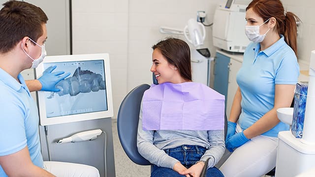
Intraoral scanners are handheld devices that capture images from inside the oral cavity. They use the same structured light technology as dental 3D scanners, photographing light distortion from the patient’s teeth and gums and generating a precise 3D image in just a few minutes. They are commonly used for indirect restorations, prosthetics and orthodontics, where they replace traditional physical impressions. According to a survey by the American Dental Association (ADA), 55% of dentists believe intraoral scanners are the number-one technology that would revolutionize their practice.
CBCT scanners
CBCT, or cone beam computed tomography, uses X-rays to create 3D images of the teeth and the hard and soft tissues of the oral cavity, as well as the nerves, muscles and bones of the head, neck and jaw. The scanner rotates around the patient from all angles, taking approximately 150-200 images, and then combines the images to construct a detailed, high-resolution 3D representation. Since CBCT provides such great depth of information, it is a valuable adjunctive tool for identifying and diagnosing oral disease, and for accurately visualizing the patient’s anatomy in preparation for more complex treatments.
Incorporating scanners into your practice
An advantage all of these scanners share is the ability to simplify and speed up various workflows within the dental practice. Here are just some of the various ways you can make use of this revolutionary technology.
1. Impressions
Many patients find the traditional method of taking physical impressions using trays to be uncomfortable and unpleasant. It can be especially difficult for younger patients and those with developmental disabilities or cognitive disorders. If a patient becomes distressed or uncooperative, the chair time is often prolonged and impressions may need to be repeated.
Patients often tell us that they find intraoral scans much more comfortable. They don’t have to tolerate the taste or texture of impression material, the pressure on their teeth and gums, or the long wait for the material to set. Dental professionals benefit too when patients are more comfortable, and there is no need to take up physical storage space or to spend time creating models or waiting for lab returns. As intraoral scans can be checked for accuracy and retaken in minutes if necessary, both patient and dental professional do not have to worry about the need for repeat appointments.
2. CAD/CAM integration
Scanners typically upload images directly to CAD/CAD (computer-aided design/computer-aided manufacturing) software, and the dental professional can visualize the patient’s anatomy in greater detail, or examine factors like margins and occlusion. The software is then used to construct anything from crowns, inlays and veneers, to dentures, bridges and implants, depending on the particular device. The final design is sent directly to a milling machine or a 3D printer to be constructed with high precision. C
CAD/CAM is becoming so accessible that much of the design and even the manufacturing process can now be completed in the dental office. Where you may have once waited weeks for restorations to come back from the lab, the entire process can be completed in-house for a fraction of the time. Alternatively, rapid digital transfer of the file to an off-site location saves time, the item can be precisely constructed in a short time and returned.
3. Oral hygiene
It can sometimes be challenging to convey to patients the importance of oral hygiene and encourage them to take preventative/corrective action. We can make use of diagrams, models and verbal explanations, but the reality is that these external representations of oral health can feel abstract to the patient and may fail to resonate.
Scanners help to make oral health and hygiene more concrete and personal. Intra-oral scanners, for example, can be used to quickly show the patient a visual of the plaque, calculus, or gingival recession happening in their own mouth at that very moment. CBCT scans can go even deeper, showing the patient markers of periodontal deterioration like alveolar bone loss. Scans can also be repeated at subsequent recalls if needed, allowing you to demonstrate any further decline (or progress!) to your patient.
As noted in Today’s RDH, patients are much more inclined to take ownership of their oral hygiene when they can see the consequences of not doing so in their own mouths. They’re also much more motivated to continue their efforts when they can see that their hard work is paying off. And when those primary barriers are overcome, they tend to be more motivated to find ways to overcome other barriers like finances.
4. Orthodontic assessment
Finally, scanners can be valuable tools for assessing the need for orthodontic treatment, generating revenue for the practice while improving patient outcomes and satisfaction. You can use scanner-generated images to quickly evaluate malocclusion and crowding; detect occlusal wear or trauma and related problems; and monitor orthodontic relapse. Scanners are used for capturing images for clear aligners, a popular choice in orthodontic treatment, and during treatment to monitor progress.


