-
-

ADULT ORTHODONTICS
Should You Use Mouthwash Before or After Brushing?Brushing and flossing are the foundation of a good oral hygiene routine, but mouthwash can also be a useful addition...

SELECTING DENTAL PRODUCTS
Soft Vs. Hard Toothbrush: Which One Should You Use?The toothbrush has come a long way. As the American Dental Association (ADA) notes...
-
Science & Innovation
- Oral Health and Dental Care | Colgate®
- Oral Health
- X-Ray Safety


Less Radiation for Safer X-Rays
Cone-Beam CT: 3-D Images, More Radiation
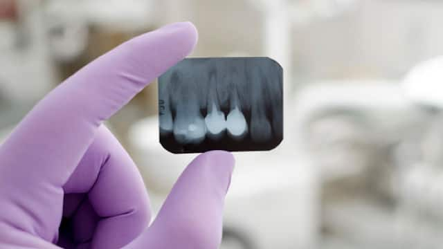
The benefits of X-rays are well known: They help dentists diagnose common problems such as cavities, gum disease and some types of infections. X-rays allow dentists to see inside a tooth and beneath the gums. Without them, more disease would go unchecked. Treatment would begin later. As a result, people would have more pain and lose more teeth.
The X-rays used in dental and medical offices emit extremely small doses of radiation. However, cells can be damaged by many small doses that add up over time. That's why experts say that X-rays should be used with caution and only when necessary.
Less Radiation for Safer X-Rays
Several changes have reduced radiation exposure in dental X-rays through the years:
- Lower X-ray dose — The single most important way dentists keep their patients safe from radiation is by limiting the dose. An X-ray machine is quite large, but the X-rays come out of a small cone. This limits the rays to an area less than three inches in diameter. X-ray machines also are well shielded. Very little radiation exposure occurs beyond the diameter of the beam.
- Better film — The speed of films used for dental X-rays has been improved. This means that less exposure is needed to get the same results. Dentists who use the fastest speed film (F-Speed) limit the amount of radiation needed to obtain a good picture. Therefore, patients also are exposed to less radiation.
- Digital radiography — The use of digital X-rays reduces radiation by as much as 80%. Today more dentists are using this type of X-ray. It's estimated that as many as one-third to one-half of U.S. dentists use this technology.
- Film holders — Dental patients used to hold X-ray film in their mouths with their fingers. Those days are long gone. Now, holders keep the film in place.
- Regular inspections and licensing — State local health departments regularly check X-ray machines to make sure they are accurate and safe.
- Lead shields — Before you get X-rays, you will be covered from the neck to the knees with a lead-lined full-body apron. Sometimes you'll also have a separate neck protector, called a thyroid collar. The American Academy of Oral and Maxillofacial Radiology recommends the use of a thyroid collar on patients under age 30. Younger adults and children are at greater risk for radiation-induced thyroid cancer than older adults. These shields have been used for decades, and many states require them. Today, however, they offer more peace of mind than actual protection. That's because modern dental X-ray machines emit almost no scatter (stray) radiation.
- Limited use of X-rays — Dentists take X-rays only when they believe they are necessary for an accurate dental assessment or diagnosis.
Current guidelines say X-rays should be given only when needed to diagnose a suspected problem. As a patient, you can help increase X-ray safety. Talk to your dentist about how often you or your children need X-rays and why.
Cone-Beam CT: 3-D Images, More Radiation
In recent years, some dentists have begun using cone-beam computed tomography (CT). These machines produce three-dimensional images of the teeth and jaw bones. Cone-beam CT exposes patients to more radiation than a standard full-mouth series of X-rays or a panoramic X-ray. Therefore, cone-beam CT should be used only where it provides a clear advantage over standard X-rays.
For selecting and placing implants, cone-beam CT is appropriate. But it is not needed for diagnosing cavities or periodontal disease. Standard X-rays also are fine for most orthodontic cases. Cone-beam CT can be useful in complex cases, however, to assist with treatment planning.
© 2002- 2018 Aetna, Inc. All rights reserved.
Related Articles
A periodontal abscess is a painful gum infection caused by bacteria in deep pockets around teeth, linked to swelling, redness, and severe discomfort.


Related Products

Helping dental professionals
More professionals across the world trust Colgate. Find resources, products, and information to give your patients a healthier future


