Types Of X-Rays
minute read
![]()
X-rays are divided into two main categories, intraoral and extraoral. With intraoral X-rays, the X-ray film is inside the mouth. With extraoral X-rays, the film is outside the mouth.
Intraoral X-rays are the most common type. They give a high level of detail. These X-rays allow dentists to:
The various types of intraoral X-rays show different aspects of the teeth:
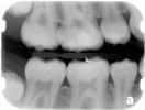
Bite-wing X-ray
highlight the crowns of the back teeth. Dentists take one or two bite-wing X-rays on each side of the mouth. Each X-ray shows the upper and lower molars (back teeth) and bicuspids (teeth in front of the molars). These X-rays are called "bite-wings" because you bite down on a wing-shaped device that holds the film in place while the X-ray is taken. These X-rays help dentists find decay between back teeth.
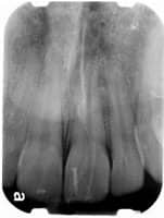
Periapical X-ray
highlight only one or two teeth at a time. A periapical X-ray looks similar to a bite-wing X-ray. However, it shows the entire length of each tooth, from crown to root.
Depending on your oral health and dental history your dentist may recommend a full-mouth radiographic surveya, or FMX. This includes every tooth, from crown to root to supporting structures. They are X-rayed using both bitewing and periapical radiographs.
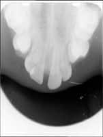
Occlusal X-ray
are larger than most X-rays. They highlight tooth development and placement in children. Each X-ray shows nearly the full arch of teeth in either the upper or lower jaw.
Extraoral X-rays are made with the film outside the mouth. These can be considered the "big picture" X-rays. They show teeth, but they also provide information on the jaw and skull. Extraoral radiographs are used to:
Extraoral X-rays are less detailed than intraoral X-rays. For this reason, they are usually not used for detecting cavities or flaws in individual teeth.
Panoramic X-rays show the entire mouth on a single X-ray. They include all teeth on both upper and lower jaws. This type of X-ray requires a special machine. The tube head that emits the X-rays circles behind your head while the film circles across the front. That way, the full, broad view of the jaws is captured on one film. Because the machine moves in a set path, you have to be positioned carefully. Devices attached to the X-ray machine hold your head and jaw in place. All this may look and feel intimidating, but the process is very safe. It often uses less radiation than intraoral X-rays.
Cephalometric projections are X-rays taken of the entire side of the head. They are used to look at the teeth in relation to the jaw and the person's profile. Orthodontists use cephalometric projections to determine the best type of orthodontic treatment.
Cone-beam computed tomography (CT) provides three-dimensional images. You stand or sit while the machine rotates around your head. The beam is cone-shaped, instead of fan-shaped as in a standard medical CT. A cone-beam scan uses less radiation than a medical CT scan but far more than any standard dental X-ray. The cone-beam CT is particularly useful for dental implant selection and placement.
Standard computed tomography (CT) usually must be done in a radiologist's office or a hospital. Typically, you will lie down while the image is taken. The radiation exposure is higher for this type of CT than for a cone-beam CT. A standard CT scan may be done to determine size and placement location for implants.
Digital radiographs are one of the newest X-ray techniques. Standard X-ray film is replaced with a flat electronic pad or sensor. The image goes into a computer, where it can be viewed on a screen, stored or printed out. Digital X-rays taken at different times can be compared using a process that highlights differences between the images. Tiny changes therefore can be caught earlier. Used properly, digital X-rays use about half the radiation of conventional film.
© 2002- 2018 Aetna, Inc. All rights reserved.