TMJ Anatomy: What To Know
January 9, 2023.
min read

The mandible, also known as the lower jaw, has two vertical extensions from each side, with a condyle at the top. The condyle can vary in shape and symmetry. The TMJ is where the condyles meet the temporal bone of the skull. A fibrous disc, called the articular disc, cushions the space between these two bones and keeps them from touching. A synovial membrane, synovial fluid, blood vessels, and nerves, as well as connective tissue, are found in the area behind the articular disc. Also, three ligaments help stabilize the joint, prevent dislocation, and support the mandibular bone weight.
Whenever you open and close your mouth, move your jaw forward, backward, or side-to-side, you are using the TMJ. This complex joint works with muscles of mastication to produce sliding and hinging motion.
According to the National Institute of Dental Craniofacial Research, almost 10 million Americans experience TMJ problems. The cause of this isn't always clear. Still, trauma to the jaw from injury, long-term teeth grinding, muscle spasms from stress, misaligned teeth, or some form of arthritis may result in TMJ disorders or TMD. Some of the signs and symptoms include popping, clicking, pain in the jaw joints, jaw locking, headaches, dizziness, earaches, neck pain, and even numbness in the fingers. Although TMJ dysfunctions tend to resolve themselves with little or no treatment, you should always visit your dentist to experience any of these symptoms.
Knowing more about how your temporomandibular joint functions may help you better understand how you can keep it in good working order. Like breathing for a count of five, relaxation techniques can help reduce stress and alleviate teeth grinding. Eating soft foods can also help to ease the tension in your TMJ. Lastly, regular dentist appointments can assist in the early detection of TMJ disorders.
This article is intended to promote understanding of and knowledge about general oral health topics. It is not intended to be a substitute for professional advice, diagnosis or treatment. Always seek the advice of your dentist or other qualified healthcare provider with any questions you may have regarding a medical condition or treatment.
ORAL HEALTH QUIZ
Take our Oral Health assessment to get the most from your oral care routine
ORAL HEALTH QUIZ
Take our Oral Health assessment to get the most from your oral care routine

Temporomandibular disorder
TMJ Arthralgia Symptoms and Treatment OptionsTMJ arthralgia is a condition within TMD that refers to pain and inflammation inside your joints. Learn more about the symptoms and treatment, here.

Temporomandibular disorder
Neuromuscular Dentistry: A DefinitionWhat is neuromuscular dentistry? Check out this great article to learn more about this specialty and when this type of dental treatment is necessary.

Temporomandibular disorder
Can Essential Oils Relieve TMJ Jaw Pain?Essential oils are used to treat a range of maladies. Learn what the science says and how TMJ essential oils may help relieve your jaw pain, here.

Temporomandibular disorder
Could Your Lateral Pterygoid Muscle Be Causing TMJ Pain?The lateral pterygoid muscle is one of the four muscles of mastication and is responsible for moving your jaw. Learn how it relates to the TMJ.
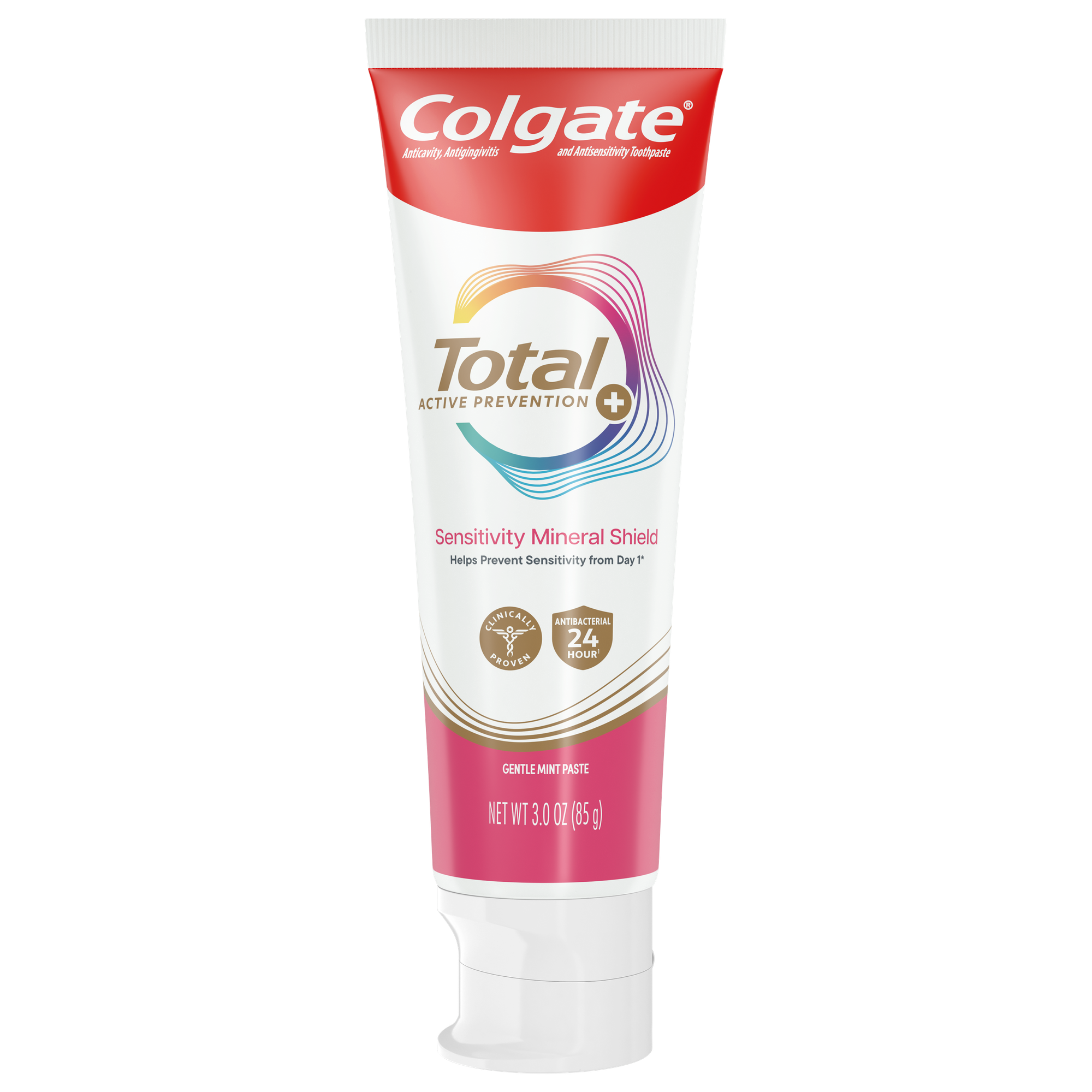
Help prevent sensitivity from day 1* with Colgate Total Active Prevention Sensitivity Mineral Shield Mint Toothpaste. This Colgate Total Sensitivity Toothpaste with Fluoride is a patented antisensitivity toothpaste that fights the root cause** of gingivitis, plaque, tartar, cavities, sensitivity, bad breath, weak enamel, and stains. Colgate Total Sensitivity Mineral Shield Toothpaste helps prevent tooth sensitivity by creating a mineral shield around sensitive spots on your teeth.
*with twice daily use
**via protection against bacteria and dietary exposures, with daily brushing
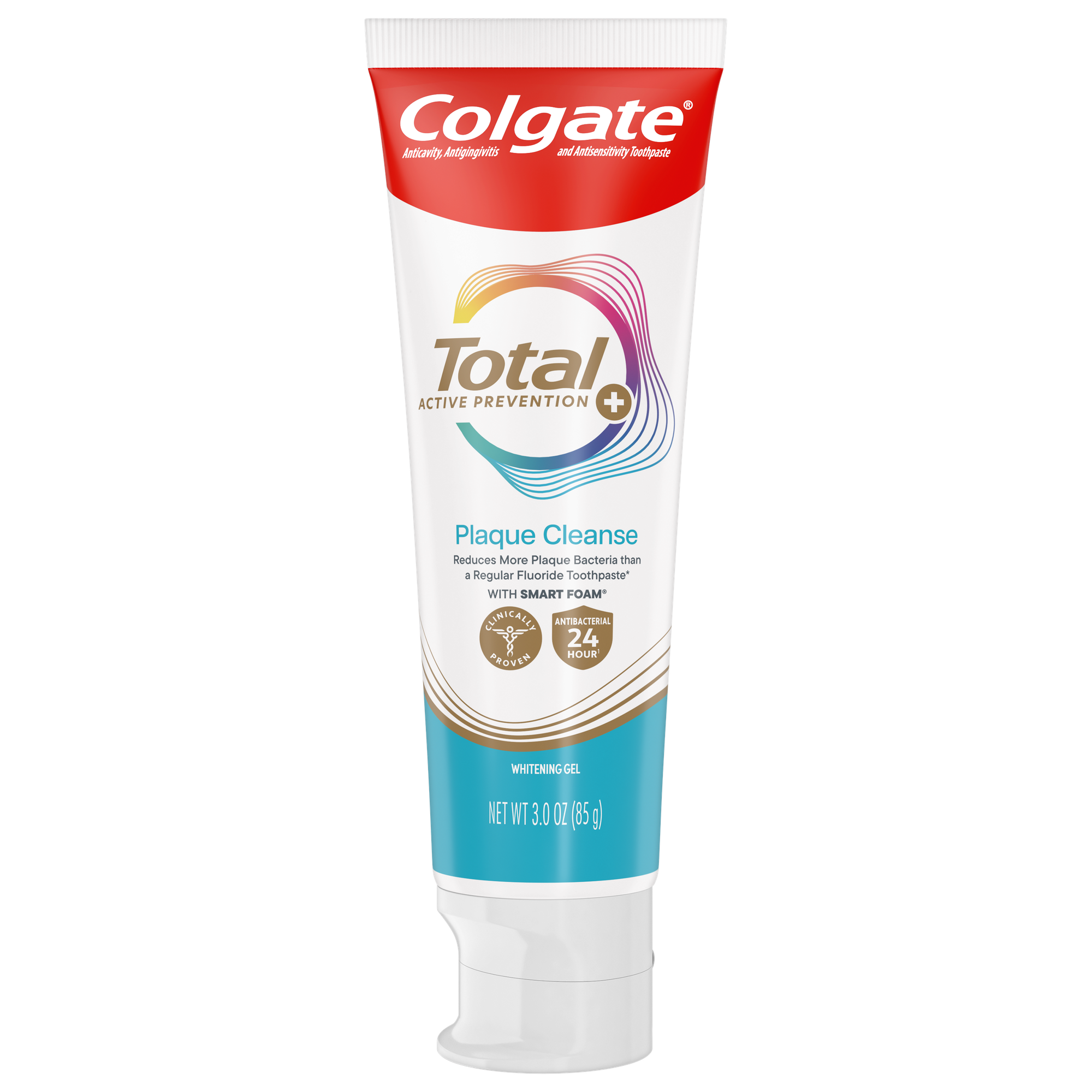
Help promote healthy gums using Colgate Total Active Prevention Plaque Cleanse Whitening Toothpaste Gel. This patented tartar prevention toothpaste fights the root cause* of gingivitis, plaque, tartar, cavities, sensitivity, bad breath, weak enamel, and stains and is 2x more effective*** at fighting bacteria, the root cause of oral health problems like cavities and gingivitis.
*via protection against bacteria and dietary exposures, with daily brushing
***via reduction of bacteria vs. non-antibacterial fluoride toothpaste with 2x daily brushing and 4 weeks use
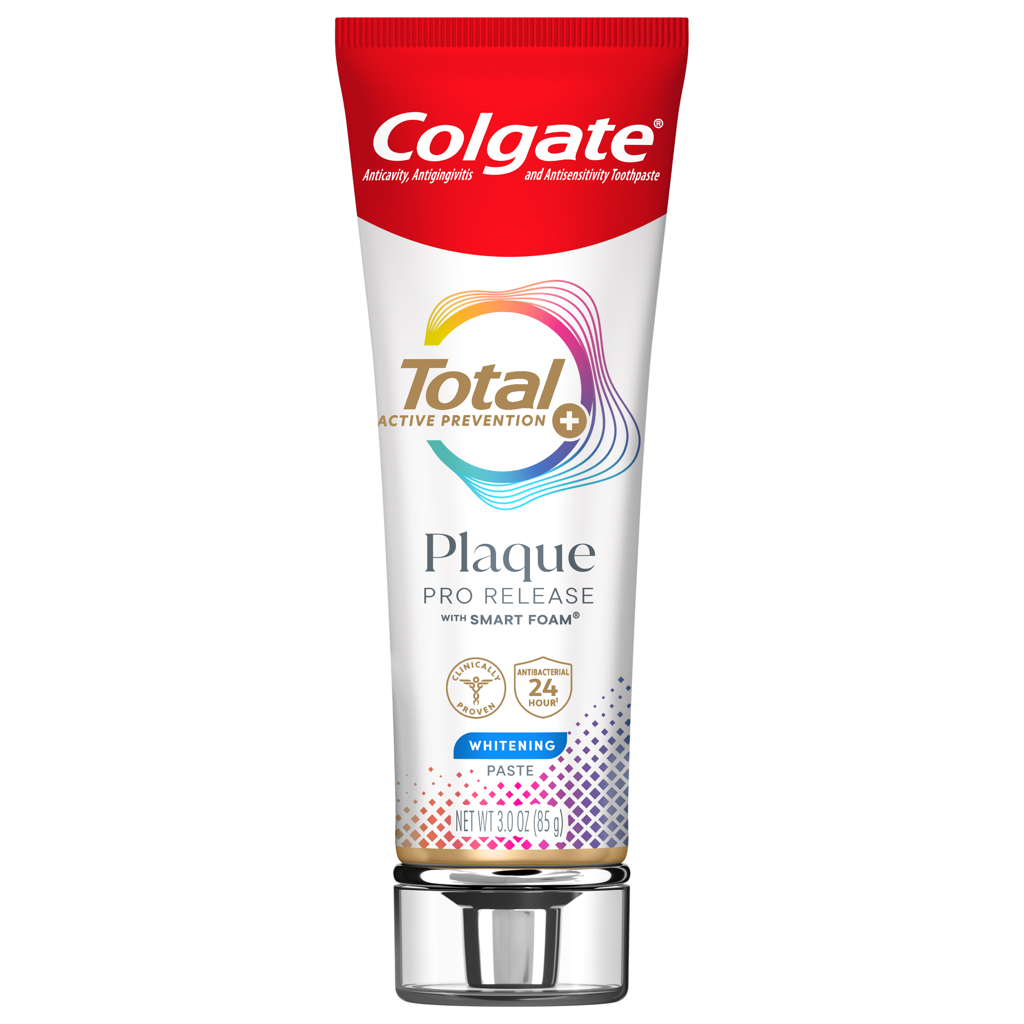
Colgate Total Plaque Pro Release Whitening Toothpaste dissolves and lifts away gum harming plaque with daily brushing.
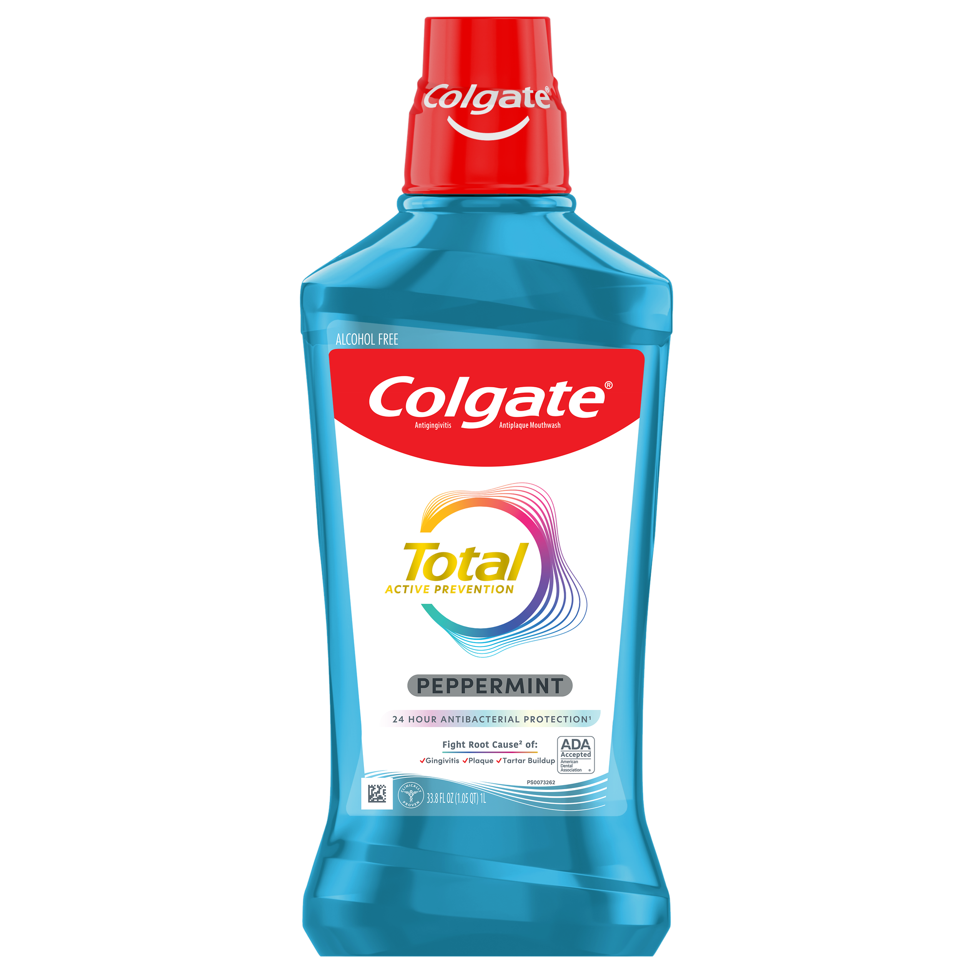
Colgate Total Alcohol Free* Peppermint Mouthwash delivers 24-hour protection** against bacteria.
