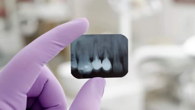
X-rays are divided into two main categories, intraoral and extraoral. Intraoral is an X-ray that is taken inside the mouth. An extraoral X-ray is taken outside of the mouth.
Intraoral X-rays are the most common type of radiograph taken in dentistry. They give a high level of detail of the tooth, bone and supporting tissues of the mouth. These X-rays allow dentists to:
- Find cavities
- Look at the tooth roots
- Check the health of the bony area around the tooth
- Determine if periodontal disease is an oral care issue
- See the status of developing teeth
- Otherwise, monitor good tooth health through prevention
Understanding
The benefits of X-rays are well known: They help dentists diagnose common problems, such as cavities, gum disease and some types of infections. Radiographs allow dentists to see inside a tooth and beneath the gums to assess the health of the bone and supporting tissues that hold teeth in place.
There are a number of X-rays a dental professional can order. The type of X-ray needed will depend greatly on the type of care the patient needs to receive.
Here are some of the most common types of X-rays performed:
- Periapical
Provides a view of the entire tooth, from the crown to the bone that helps to support the tooth. - Bite-Wing
Offers a visual of both the lower and upper posterior teeth. This type of X-ray shows the dentist how these teeth touch one another (or occlude) and helps to determine if decay is present between back teeth. - Panoramic
Shows a view of the teeth, jaws, nasal area, sinuses and the joints of the jaw, and is usually taken when a patient may need orthodontic treatment or implant placement. - Occlusal
Offers a clear view of the floor of the mouth to show the bite of the upper or lower jaw. This kind of X-ray highlights children’s tooth development to show the primary (baby) and permanent (adult) teeth.
These X-rays are typically performed in the office of a dentist or dental specialist. First, a dental professional will cover you with a heavy lead apron to protect your body from the radiation. Next, the dental professional will insert a small apparatus, made of plastic, into your mouth and ask you to bite down on it - this holds the X-ray film in place. The technician will then proceed to take an X-ray picture of the targeted area. This process is pain-free and will be repeated until images have been obtained for your entire mouth. The use of digital X-rays provides significantly less radiation to the dental patient and is convenient and time saving for the dental practice.
Planning
Dental X-rays are very safe and expose you or your child to a minimal amount of radiation. When all standard safety precautions are taken, today's X-ray equipment is able to eliminate unnecessary radiation and allows the dentist to focus the X-ray beam on a specific part of the mouth. High-speed film enables the dentist to reduce the amount of radiation the patient receives. A lead body apron covers the body from the neck to the knees and protects the body from stray radiation.
Oral Care Center articles are reviewed by an oral health medical professional. This information is for educational purposes only. This content is not intended to be a substitute for professional medical advice, diagnosis or treatment. Always seek the advice of your dentist, physician or other qualified healthcare provider.
ORAL HEALTH QUIZ
What's behind your smile?
Take our Oral Health assessment to get the most from your oral care routine
ORAL HEALTH QUIZ
What's behind your smile?
Take our Oral Health assessment to get the most from your oral care routine


