Types of X-rays
January 9, 2023.
min read

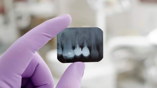
Intraoral Radiographs are the most common type of dental X-rays you’ll encounter during a routine dental exam. Your dentist is looking for cavities and checking the status of developing teeth. These radiographs also give your dentist the ability to view tooth roots, check the health of the bone and even diagnose periodontal disease. The different types of intraoral X-rays show different aspects of the teeth.
Like the first part of the name suggests, extraoral X-rays are made with the film outside the mouth. This type of X-ray still shows the teeth but can also provide important information about the jaw and skull. Think of these X-rays like the “big picture” of oral health, they are used to see how everything comes together.
Digital radiographs use a flat electronic pad or sensor that runs the images through a computer instead of X-ray film and provide the lowest level of radiation. According to MedlinePlus the amount of radiation you’re exposed to with these types of X-rays is much less than a traditional X-ray.
Yes! According to the American Dental Association, dental X-ray exams are safe. Wondering if you’ll have X-rays at your upcoming appointment? Depending on factors like age, risk for disease, present oral health, and if you’re a new patient, X-rays may be part of your exam. As with all X-rays, the procedure is quick and painless.
Oral Care Center articles are reviewed by an oral health medical professional. This information is for educational purposes only. This content is not intended to be a substitute for professional medical advice, diagnosis or treatment. Always seek the advice of your dentist, physician or other qualified healthcare provider.
ORAL HEALTH QUIZ
Take our Oral Health assessment to get the most from your oral care routine
ORAL HEALTH QUIZ
Take our Oral Health assessment to get the most from your oral care routine

Thanks to dental x-rays dentists can accurately diagnose and treat dental problems before they become more serious. Learn more here.
If you're on a tight budget you may worry if the cost of dental X-rays is worth it, even if your dentist does recommend it. Find out more about costs here.

Unlike A traditional radiograph, a panoramic dental x-ray creates a single image of the entire mouth including upper and lower jaws, TMJ joints, teeth, and more.
X-Rays are divided into two main categories, intraoral and extraoral. Find out more about intraoral and extraoral radiographs, here.
Help prevent sensitivity from day 1* with Colgate Total Active Prevention Sensitivity Mineral Shield Mint Toothpaste.
*with twice daily use
Help promote healthy gums using Colgate Total Active Prevention Plaque Cleanse Whitening Toothpaste Gel. This patented tartar prevention toothpaste fights the root cause* of gingivitis, plaque, tartar, cavities, sensitivity, bad breath, weak enamel, and stains and is 2x more effective*** at fighting bacteria, the root cause of oral health problems like cavities and gingivitis.
*via protection against bacteria and dietary exposures, with daily brushing
***via reduction of bacteria vs. non-antibacterial fluoride toothpaste with 2x daily brushing and 4 weeks use
Colgate Total Plaque Pro Release Whitening Toothpaste dissolves and lifts away gum harming plaque with daily brushing.

Colgate Total Alcohol Free* Peppermint Mouthwash delivers 24-hour protection** against bacteria.
